|

info@analyticalcontrols.com

317-841-0458

317-841-3186

Analytical Control Systems, Inc.
All WebPages' contents are © 2002-2004 Analytical Control Systems, Inc.
Many of Analytical Control Systems, Inc. products are covered by US and
foreign patents or patents pending.
| |
Platelet Derived Growth Factor and Hemostatin™
Determination of Platelet Derived Growth Factor
Drs. Xiangdong Cui, M.D. and Dayong Gao, Ph.D. performed a study of
Hemostatin’s™ effect on the release of growth factors from platelet activation
at the Department of Mechanical Engineering and Center for Biomedical Engineering at the
University of Kentucky.
Introduction: Platelet Derived Growth Factor (PDGF) is a potent
mitogen/chemoattractant for connective tissue and plays an important role in wound healing
and angiogenesis. The target cells for PDGF include mammalian cells, such as fibroblasts,
endothelial cells, and smooth muscle cells. Our present experiment is to observe where the
PDGF exists, how effective PDGF is for cell growth and to observe some related phenomenon
about propyl gallate (PG) (Hemostatin™).
Materials and Methods:
1. Preparation of the samples:
a. Obtain platelets from the blood bank of UK hospital (fresh, 0 days old);
b. Adjust platelet count to 250,000/mm3 (with TB buffer);
c. Add 1 ml of PG reagent (1:1, or 1:5) and stir for 5 min.;
d. Divide the sample into supernatant and aggregates and freeze at –70°C for later use;
e. Take part of the platelet sample (2-3 ml), centrifuge to remove all of the platelets
and get platelet poor plasma (PPP). Freeze at –70°C for
later use.
2. Order the PDGF and relative chemicals from Promega:
Platelet Derived Growth Factor PDGF(AB), Human Recombinant; PDGF(AA), Human
Recombinant; PDGF(BB), Human Recombinant; Transforming Growth Factor-alpha, Human
Recombinant (rh TGF-a); Fibroblast Growth Factor, Basic, Human Recombinant (rh FGF,
Basic); Fibroblast Growth Factor, Acidic, Human Recombinant (rh FGF, Acidic);
3. Order Balb/3T3, Clone A31 cells from ATCC:
a. One vial, seed and culture them for experimental use. The medium: DMEM/F12
4. CellTiter 96™ Non-radioactive Cell Proliferation/Cytotoxicity Assay.
a. Three days prior to performing this assay, seed 1-2X10⁵ 3T3 cells to t-75
flask in F12/DMEM supplemented with 10% calf serum;
b. The column 1 was set as the negative control;
c. Add 100 ml of the cell suspension (containing 10,000 cells) to each well of the 96
well plate and return to the incubator for 24 hours;
d. The next day, add 20 ml/well reagent and then continue to culture for 48 hours;
e. Add 15 ml/well of CellTiter 96™ Dye Solution and
incubate the plate for 4 hours;
f. Add 10 ml/well of CellTiter 96™ Solubilization/Stop
Solution and keep it at 37°C in an incubator overnight;
g. Record the absorbance at 570 nm using an Elisa plate reader. Use of a reference
wavelength is optional (630 nm).
h. Plot the corrected absorbance at 570 nm (y– axis) versus ng/ml of growth factor
(x- axis).
We found that PG can induce the platelets to aggregate and release PDGF, and the latter
can enhance growth of cells. But, it may inhibit the condition of cells. In order to
investigate this phenomenon, we did other experiments for the dose dependent test.
Results:
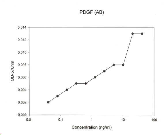
PDGF(AB)
Supernatant 12.5 ng/ml
Platelet debris 1.3 ng/ml
PPP 0 ng/ml
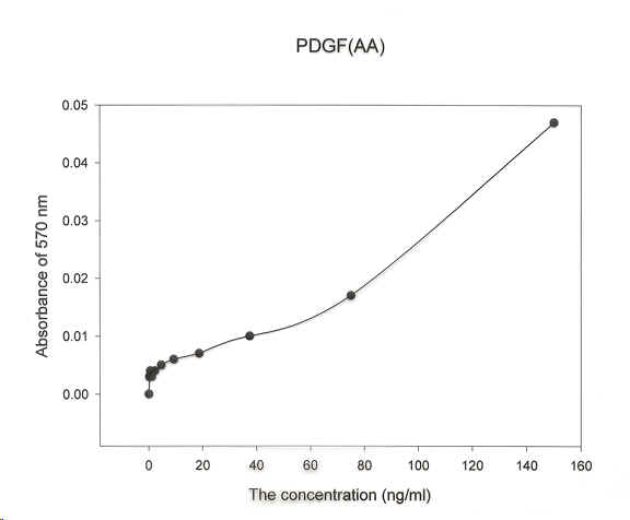
PDGF(AA)
Supernatant 18.1 ng/ml
Platelet debris 1.2 ng/ml
PPP 0 ng/ml
PDGF(BB)
Supernatant 11.6 ng/ml
Platelet debris 2.1 ng/ml
PPP 0 ng/ml
Rh FGF- Basic
Supernatant 7.5 ng/ml
Platelet debris 0 ng/ml
PPP 5.3 ng/ml
Rh FGF- Acidic
Supernatant 1.2 ng/ml
Platelet debris 0.7 ng/ml
PPP 6.8 ng/ml
TGF-a
Supernatant 1.4 ng/ml
Platelet debris 0 ng/ml
PPP 50.8 ng/ml
According to our experiments, the preliminary conclusions are that:
- PG is a potent reagent for platelet aggregation;
- Platelets can be thoroughly activated and broken and release PDGF by PG;
- Most of the PDGF is contained in the supernatant, with a few of them in the debris of
the platelets;
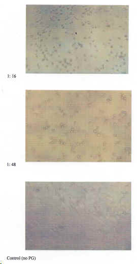
- The mixed PG and PDGF solution can enhance growth of 3T3 cells in vitro;
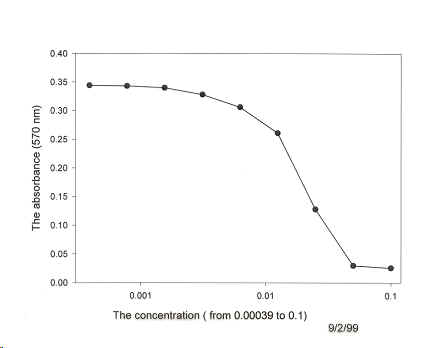
- In the relatively high concentrations of PG only, PG could inhibit the proliferation of
the cells;
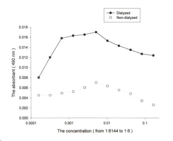
- When comparing the dialyzed and non-dialyzed platelets freeze dried crystal, the
efficiency of dialyzed is better.
|